Introduction
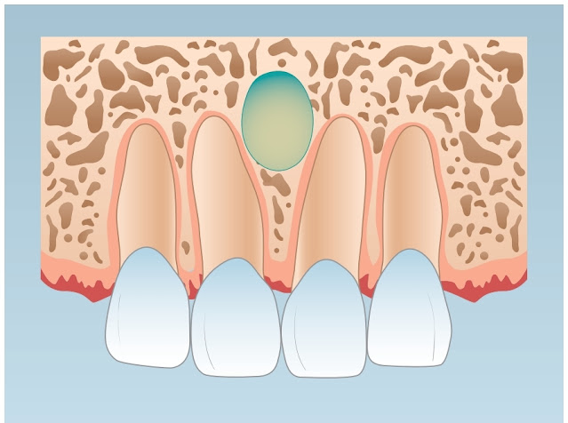
An intraosseous developmental cyst of the midline of the anterior palate, derived from islands of epithelium remaining after the closure of the embryonic nasopalatine duct.
It is also known as incisive canal cyst.
Clinical feature
- These are mostly intraosseous.
- But sometimes it can be present extraosseous entirely in soft tissue.
- They are usually painless and detected on routine radiograph.
- But can also cause pain and inflammation and present.

Radiograph
- In edentulous patients, they do not appear properly.
- But in a dentate patient, it appears oval, heart-shaped, or pear-shaped.
- Present between roots of central incisors.


Histopathology
- Lining epithelium can be ciliated columnar, cuboidal, or stratified squamous.
- If inflammation is present it has lymphocytes or plasma cells.
- The capsule has nerves and vessels which are contents of the incisive canal.


No comments:
Post a Comment