Wednesday, 29 June 2022

Dental caries and its classification slideshare ppt

Dental caries

It is a multifaceted disease involving teeth host factors of saliva and microflora and the external factors of diet. In it, unique strains of bacteria accumulate on the enamel surface where they release acidic and proteolytic products that demineralize the surface and digest the organic matrix.

Types of caries

  1. Pit and fissure caries
  2. Smooth surface caries
  3. Cemental caries
  4. Recurrent caries

Acute caries

  • Also known as ramapant caries
  • Progress at a high rate
  • Common in children and young adults
  • Teeth have large pulpal chambers and short and wide tubules with slight sclerosis
  • Patient has diet high in refined carbohydrate

Chronic caries

  • Common in old people
  • Teeth have small pulp chambers
  • Less tubular dentin is present
  • More sclerosis is present
  • There is secondary and tertiary dentin
  • Less severe pain  
For more watch 

Tuesday, 28 June 2022

Dermoid vs epidermoid cyst / histopathology, clinical and treatment

Dermoid Cyst

A cyst of the midline of the upper neck of the anterior floor of the mouth of young patients derived from remnants of embryonic skin consisting of a lumen lined by a keratinizing stratified squamous epithelium and containing one or more skin appendages such as hair, sweat, or sebaceous glands.

  • It is an example of cystic teratoma
  • Derived from germinal epithelium entrapped during embryonic development

Clinical features

  • It is present in young adults
  • Present in the upper neck or floor of the mouth
  • Present as painless swelling
  • It has doughy consistency
  • It is 2cm or less in diameter

Histopathological features

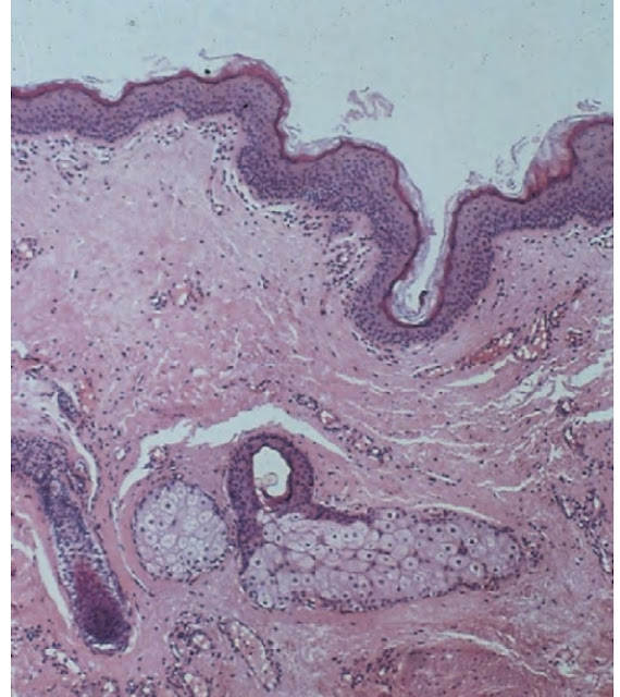
  • The lining is orthokeratinized squamous epithelium.
  • Exhibiting a variable number of dermal appendages such as hair follicles sebaceous glands and erector pili muscles.
  • Lumen is filled with desquamated keratin sebum and hair shafts.
  • There is a sorrounding zone of compressed connective tissue.
  • Generally free of inflammation.

Epidermoid cyst

  • Same as dermoid cyst but skin appendages are absent.
  • Treatment is surgical enucleation. 
For more watch here

Friday, 24 June 2022

Thyroglossal tract cyst / histopathology, and clinical features slideshare ppt

Introduction

A cyst located above the thyroid gland and beneath the base of the tongue with a lumen lined by a mixture of epithelial cell types derived from remnants of the embryonic thyroglossal tract and often containing thyroid tissue in the capsule.

  • The tract extends from the foramen caecum on the mid dorsum of the tongue to the thyroid gland. 
  • 70 to 80% occur below the hyoid bone.

Clinical features

  • Present in children and young adults.
  • Asymptomatic and slowly enlarging.
  • Mobile moves upwards with tongue protrusion.
  • Present in the anterior neck in the midline.
  • It can form a draining fistula.

Histopathology

  • Lined by stratified squamous epithelium, transition epithelium or ciliated columnar epithelium, or its types.
  • Cyst capsule has thyroid Tissue, mucus glands, sebaceous glands.
  • Carcimoma can develop occasionally.

Treatment

  • Operation is Sistrunk operation.
  • All tissues involved in the tract from the base of the tongue to the thyroid gland including the middle portion of the hyoid bone.
For more watch here

Thursday, 23 June 2022

Nasolabial cyst / histopathology, clinical and radiographic features slideshare ppt

Nasolabial cyst

It is a developmental Cyst of soft tissue located in the anterior mucobuccal fold beneath the ala of the nose and most likely developed from remnants of an inferior portion of nasolacrimal duct.

It is also known as nasoalveolar cyst or klestadt cyst.

Clinical features

  1. It is present entirely in soft tissue in the anterior maxillary vestibule 
  2. Below ala of the nose deep in the nasolabial crease
  3. It is unilateral sometimes bilateral painless soft tissue swelling
  4. Nasolabial crease appears to be absent
  5. If we retract the upper lip properly we can see cyst swelling in the vestibule
  6. Cyst occurs in 4th to 5th decade of life

Radiographic features

  1. Since cyst is present in soft tissue so it is not visible in a radiograph
  2. However contrast can be used for better visualization
  3. Pressure-induced saucerization of underlying bone of maxilla can be seen

Histopathology

  1. The epithelium is pseudostratified columnar with a variable number of goblet cells (mucous cells) lined by cuboidal cells
  2.  A narrow zone of dense homogeneous fibrous tissue is seen adjacent to the cyst lining
For more watch here

Monday, 20 June 2022

Cementum and composition of cementum slideshare ppt

Cementum

It is hard, avascular connective tissue which covers that covers the roots of teeth. 

Development of cementum

It is divided into two stages

1. Prefunctional stage

It occurs throughout root formation.

2. Functional stage

It starts when the tooth is in occlusion and continues throughout life. 

Composition of cementum

Cementum consists of both inorganic and organic components

Inorganic component

Cementum consists of 45-50% hydroxyapatite crystal by weight.

Organic Component

It consists of collagen and non-collagenous matrix protein

  • Collagen type I forms 90% of organic constituents in cellular cementum. 
  • Collagen type III and XII and a trace amount of collagen type V, VI, and XIV are also found in cementum.
  • Non-collagenous matrix protein consists of alkaline phosphatase, bone sialoprotein, dentin matrix protein 1, dentin sialoprotein, fibronectin, osteocalcin, osteonectin, and several growth factors.
For more watch here

Sunday, 19 June 2022

Nasopalatine duct cyst / Histopatholgy, clinical and radiographic feature slideshare ppt

Introduction

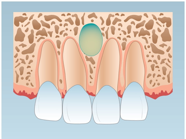
An intraosseous developmental cyst of the midline of the anterior palate, derived from islands of epithelium remaining after the closure of the embryonic nasopalatine duct.

It is also known as incisive canal cyst.

Clinical feature

  1. These are mostly intraosseous.
  2. But sometimes it can be present extraosseous entirely in soft tissue.
  3. They are usually painless and detected on routine radiograph.
  4. But can also cause pain and inflammation and present.

Radiograph

  1. In edentulous patients, they do not appear properly.
  2. But in a dentate patient, it appears oval, heart-shaped, or pear-shaped.
  3. Present between roots of central incisors.

Histopathology

  1. Lining epithelium can be ciliated columnar, cuboidal, or stratified squamous.
  2. If inflammation is present it has lymphocytes or plasma cells.
  3. The capsule has nerves and vessels which are contents of the incisive canal. 
For more watch here

Saturday, 18 June 2022

Periodontium components / Parts of periodontium slideshare ppt

 The periodontium is defined as those tissues supporting and investing the tooth.

Components of periodontium

  1. Cementum
  2. Periodontal ligaments
  3. Bone lining the alveolus
  4. Gingiva facing the tooth

1. Cementum

  • The cementum is a hard, avascular connective tissue that covers the roots of teeth.
  • Cementum is classified according to the presence or absence of cells within its matrix and the origin of the collagen fibers of the matrix. 

2. Periodontal ligaments

  • The PDL is that soft, specialized connective tissue situated between the cementum covering the root of the tooth and the bone-forming the socket wall.
  •  It attaches a tooth to the alveolar bone.

3. Gingiva

  • It is the soft, pink tissue that surrounds and protects the bottom of the teeth where they enter the jawbone.

4. Alveolar process

  • The alveolar process is one of the jaws containing the sockets (alveoli) for the teeth. 

Functions of periodontium

  1. It provides support to the tooth.
  2. It protects the tooth against oral microflora. 
  3. Attachment of the tooth to the bone.
For more watch here

Diagram showing Components of Periodontium


Thursday, 16 June 2022

Glandular odontogenic cyst / histopathology, radiographic and clinical features ppt slide share

 Introduction

A unilocular or multilocular odontogenic cyst derived from the dental lamina and characterized by a lining containing glandular cells lined by cuboidal or columnar cells often including mucous cells.

 Also known as Sialo-odontogenic cyst.

Clinical features

  • Most commonly occur in the mandible. 
  •  It has high growth potential. 
  •  It can reoccur.
  •  Other properties are similar to Lateral periodontal Cyst.

Radiographic features

  • It appears well defined unilocular or multilocular radiolucency.
  • Most commonly occur in the mandible.

Histopathology

  • It has a lining of stratified squamous epithelium.
  •  May have local thickenings plaque.
  •  Small number of microcyst or glandular structures within epithelium.
  •  Single layer of cuboidal or columnar cell lining glandular structure.
  • It is mucaramine and PAS +ve.

Treatment

  • By surgical enucleation.

For more watch here


Pulp stones / denticles

Pulp stones or denticles are discrete masses that have a calcium phosphate ratio comparable to that of dentin. It may be singular or multiple.

Histology

They consist of concentric layers of mineralized tissue formed by accretion around blood thrombi, dead cells, or collagen.

Classification of pulp stones

There are several ways of classification of pulp stone.

1.

a) True pulp stones.

b) False pulp stones.

2.

a) Free pulp stones.

b) Attached pulp stones.

Clinical significance

  • Asymptomatic.
  • Large enough to be detected radiographically.
  • Problematic during endodontic therapy.
For more watch here
Lecture notes by Dr.Bala

Tuesday, 14 June 2022

Lateral periodontal cyst and ginival cyst of adult

 Introduction of lateral periodontal cyst

A slow-growing non-expansile odontogenic cyst derived from the rest of dental lamina exhibiting a lining of 2 to 3 layers of cuboidal cells and distinctive focal thickenings (plaques).

  • Sometimes polycystic variant is present and known as a Botryoid cyst.

Clinical features

  1. Most commonly present between premolars or between the roots of the incisor. 
  2. The mean age of occurrence is 50 years.
  3. Usually 1cm in diameter
  4. It has a slow expansile rate.
  5. Recurrence is rare.
  6. Associated teeth are vital. 

Radiographic features

  1. It appears well defined.
  2. Unilocular.
  3. Delicately corticated radiolucency. 
  4. Present between roots of vital teeth.

Histopathology

  1. The lining epithelium is non-keratinized with two to three layers of cuboidal cells.
  2. But there are focal thickenings known as plaques.
  3. The cell layer has glycogen-rich clear cells.

Treatment

Treatment is surgical enucleation. 

Gingival cyst of adult

  • It is considered the extraosseous counterpart of lateral periodontal cyst
  • All the features are the same including histology and time of occurrence.
  • It is firm yet compressible fluid-filled swelling. 
  • On a radiograph, it is not apparent but sometimes it can cause saucerization of the underlying bone.
For more watch 

Monday, 13 June 2022

Age changes in dentin-pulp complex

 Following changes appear in the dentin-pulp complex with time.

  1. Decrease volume of the pulp chamber and root canal due to continuous deposition of dentin.
  2. Decreased vascular supply to pulp.
  3. Reduction in the number of cells. The number of cells becomes half between ages 20 to 70.
  4. Loss of myelinated and unmyelinated axons leads to a reduction in the sensitivity of dentin.
  5. Increase in dead tracks and sclerotic dentin.
  6. The appearance of areas of dystrophic calcification.
  7. Increased sclerotic dentin deposition leads to increased brittleness and decreased permeability.
For more watch Here
Lecture notes by Dr.Bala

Sunday, 12 June 2022

Odontogenic keratocyst / histopathology, clinical and radiographic features

 Odontogenic keratocyst ( OKC )

  • A cyst derived from the remnants of dental lamina with a biological behaviour similar to a benign neoplasm with a distinctive lining of 6 to 10 cells in thickness and that exhibit a basal cell layer of palisaded cells and a surface of corrugated  parakeratin.
  • Odontogenic keratocysts (OKC) are ususally present intraosseous but extraosseous OKC sometimes also present termed as peripheral OKC.

Clinical features

  • Occurs in wide age span 1st decade to 8th decade of life but most commonly present in 2nd and 3rd decade of life.
  • Most commonly present in posterior body of the ramus. 
  • Sometimes it resemble dentigerous cyst but is not limited to CEJ.
  •  It has remarkable growth potential resulting in massive bone destruction.
  •  Reoccurrence rate is 25 to 60% similar to ameloblastoma.

Radiographic features

  •  Appear well defined solitary lesion with smooth or scalloped margins or as multilocular radiolucency exhibiting thin corticated margin. 

Histopathological features

  •  Thin uniform Lining of perakeratinized squamous epithelium usually 6 to 10 cell layer thick.
  •  A palisaded layer of columnar or cuboidal basal cells.
  •  A corrugated rippled layer of parakeratin on its luminal surface.
  •  A lack of rete pegs.
  •  Cystic lumen contain desquamated parakeratin. 
  •  Also has satellite cyst.
  •  Parakeratin has corrugated appearence. 

Why OKC has high recurrence?

  • It has satellite cyst.
  •  There is focal seperation of epithelial lining from connective tissue.
  •  Lack of rete pegs.
  •  Parakeratinzed OKC has higher recurrence rate than orthokeratinized vairiant which is 5%.

Nevoid basal cell carcinoma

Features of nevoid basal cell carcinoma:

  • Multiple odontogenic keratocysts of jaws
  • Multiple basal cell carcinoma of skin
  • Bifid ribs
  • Calcification of the falx cerebri
  • Palmar and plantar dyskeratosis
  • Multiple epidermoid cysts (milia) of skin
  • Frontal bossing
  • Hypertelorism
  • Ovarian fibromas
  • Medulloblastoma
  • Shortened metacarpals
For more watch here
  • Forhead exhibiting frontal bossing with skin "milia"

    Calcification of falx cerebri

Saturday, 11 June 2022

Dentin hypersensitivity / Theories of dentin sensitivity

Dentin Hypersensitivity

It is characterized by short, sharp pain arising from exposed dentin in response to stimuli like thermal, evaporative, tactile or chemical.

Causes / Etiology of dentin hypersensitivity

1. Erosion 

2. Abrasion

3. Parafunctional habits

4. Occlusal wear

5. Gingival recession

6. Periodontal disease

Theories regarding dentin sensitivity

  •  Direct Innvervation Theory
  •  Transduction / Odontoblast deformation theory
  •  Hydrodynamic theory

For more watch here

Lecture notes by Dr. Bala

Lecture notes by Dr. Bala

Lecture notes by Dr. Bala

Friday, 10 June 2022

Paradental cyst / histopathology, clinical and radiographic features

Paradental cyst 

A cyst of odontogenic origin commonly located subgingivaly on buccal aspect of  an erupted mandibular molar (bifurcation cyst) or the distal surface of a partially erupted mandibular third molar.

▪ Also known as Criag cyst.

▪ Buccal bifurcation cyst.

Clinical features

▪ Cyst is inflamed but tooth is vital is present at bifurcation area usually associated with mandibular molar which has cervical enamel projection.

▪ It can also be present distally of third molar which is anatomically impeded by ascending ramus. 

▪ Sometimes paradental cyst give rise to Garres osteomyelitis. 

Radiographic features

▪ Buccal bifurcation cyst cannot be visible on routine radiograph.

▪ But cyst present distally of third molar can be visible on periapical panoramic radiograph.

Histopathology

▪ Epithelium is non keratinized squamous eptihelium

▪ Usually infiltrated by neutrophils

▪ Connective tissue is chronically inflamed

▪ It has resemblance with dentigerous cyst

Treatment

▪ Usually treated by surgical enucleation. 

▪ Molar is often extracted during surgery.

For more watch Here

Wednesday, 8 June 2022

Dentigerous and periapical cyst / histopathology, clinical and radiographic features

Dentigerous cyst

A odontogenic cyst that surrounds the crown of impacted tooth. It is caused by fluid accumulation between reduced enamel epithelium and crown. Resulting a cyst in which crown is within lumen and roots are outside.

Clinical Feature

▪ It is usually associated with impacted canine or third molar.

▪ It is usually asymptomatic but sometime pain and inflammation is present.

▪ In arch the tooth appear to have missing one tooth.

Radiographic features

It is usually diagnosed by radiograph

▪ It appears well circumcised radiolucency sorrounding the impacted tooth.

▪ Radiolucency limited to cementoenamel junction.

▪ Sorrounding bone is corticated.

Histopathology

▪ Epithelium is  non-keratinized stratified squamous epithelium. 

▪ It is 2 to 10 cell layer thick.

▪ Inflammation can be present.

▪ It has rushton bodies.

▪ Cholesterol and hemosiderin deposit.

▪ Lipid laden macrophages.

▪ Cell layer may have mucous cells known as mucous cell mataplasia.

Importance

Since it has a malignant potential, so it can give rise to

▪ Mucoepidermoid carcimoma

▪ Ameloblastoma

▪ Squamous cell carcinoma

Eruption cyst

An odontogenic cyst with the histologic feature of a dentigerous cyst that sorrounds the tooth crown that has erupted through bone but not through soft tissue and is clinically visible as a soft fluctuant mass on the alveolar ridge.

▪ It does not form intrabony radioluency.

▪ Sometimes it ruptures forming erruption hematoma.

▪ It has variable number of ghosted cell present in cyst lumen.

More more watch here


Tuesday, 7 June 2022

Dental pulp and its histology

Dental Pulp

The soft connective tissue which is present inside the pulp cavity and supports the overlying dentin is called as pulp. It is characterized by the presence of nerve and blood vessels.

Pulp Cavity

The cavity which contains the pulp is known as pulp cavity. It is further divided into two on the basis of its location.

1. Pulp chamber

The portion of pulp cavity which is present inside the crown of the tooth. It contains the cronal pulp.

2. Root canal

The part of pulp cavity which is present in the root of the tooth is called root canal or root canal system in multirooted tooth. It contains the radicular pulp.

Histology of the Pulp

Histological examination of the pulp shows four distinct zones which are

1. The odontoblastic zone at the pulp periphery.

2. A cell-free zone of Weil beneath the odontoblasts which is prominent is the coronal pup

3. A cell-rich zone where cell density is high, as seen adjacent to the cell-free zone.

4. The pulp core which is characterized by the major vessels and nerve of the pulp.

Principal Cells of the Pulp

Cells which are present predominantly inside the pulp are

1. Odontoblasts are the most prominent cells in pulp.

2. Fibroblast are most numerous type of cells in pulp.

3.  Undifferentiated ectomesenchymal cells. 

4. Macrophages and other immunocompetent cells.

For more watch Here

Lecture notes by Dr.Bala

Lecture notes by Dr. Bala


Monday, 6 June 2022

Periapical cyst / Radiology, histopathology and clinical features

An odontogenic cyst derived from rest of malasssez proliferate in response to inflammation. It is also known as apical periodontal cyst or redicular cyst.

It is associated with devitalized tooth and form in response to pulp inflammation or necrosis.

Histopathology:

1. Epithelium is nonkeratinized stratified squamous epithelium.

2. Connective tissue contain inflammatory cells such as neutrophils.

3. Cyst has laminated hyaline bodies known as ruston bodies.

4. Cyst wall has multinucleated giant cells, cholesterol and hemosiderin.

5. Cystic lumen has proteinaceous fluid.

Formation of periapical cyst:

1. Cells of the rest of malassez proliferate in response to inflammation.

2. As cells proliferate inner cells get deficient of nutrients which are getting by duffusion.

3. So central cells undergo ischemia and liquifactive necrosis.

4. Central necrotic area creates an osmotic gradient which draws water inwards causing hydraulic pressure in cyst too expand.

Clinical Features:

1. It is associated with devitalized tooth which is its identifying feature.

2. It is more commonly present isn apical areas near canal opening.

3. It is usually 1 cm in diameter.

4. If the tooth is extracted without removing the cyst it will form Residual cyst.

Radiographic features:

1. It appears as well circumscribed corticated radiolucency at the apex of the nonvital tooth.

2. It is present laterally then appears semicirular

3. In anterior teeth is is reffered as globulomaxillary radiolucency.

Differential diagnosis:

a. Periapical granuloma.

b. Lateral periodontal cyst.

c. Odontogenic keratocyst.

d. Central giant cell granuloma.

e. Calcifying odontogenic tumor.

f. Adenamatoid odontogenic tumor.

g. Odontogenic myxoma.

For more watch here

Sunday, 5 June 2022

Interglobular dentin / Incremental growth lines / Granular layer of tomes slideshare ppt

Interglobular dentin

These are areas of hypomineralization or calcification due to failure of fusion of globular masses of mineralization.

It is a defect in mineralization and dentin architecture is normal or unchanged. 

It is most commonly seen in circumpulpal dentin.

Causes:

It is due to the deficiency of vitamin D or high fluoride level during mineralization.

Incremental Growth Lines

These represents the rhythmic pattern of organic matrix deposition of dentin.

It is deposited at the rate of 4 mm per day. 

Von Ebner Lines

Incremental lines which are at right angle to dentinal tubules are known as Von Ebner Lines

These represent 5 days deposition of dentin.

Neonatal Lines

Wide contour line found in those teeth which are mineralizing at birth.

It represents the disturbance in mineralization created by the physiologic trauma of birth.

Granular Layer of Tomes

Present in root dentin just below the cementum and extend from cementoenamel junction ( CEJ ) to root apex.

They are wider at apex of the tooth then at the CEJ.

Why Granular Layer of Tomes formed?

It is due to

1. Area of hypomineraliztion (Interglobular dentin).

2. Looped terminal portion of dentinal tubules.

3. Special arrangement of collagenous and non-collagenous proteins.

For more watch here

Lecture notes by Dr. Bala

Lecture notes by Dr. Bala

Lecture notes by Dr. Bala


Saturday, 4 June 2022

Cysts of the oral regions / Classification of odontogenic cyst

Cyst:

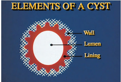
It is a pathological cavity lined by epithelium and lumen contain semisolid fluid. Cyst is mainly composed of three parts: 

1. Epithelium

2. Connective tissue capsule

3. Central  lumen

Inflammation is often present which can distort its normal morphology.

Cyst is broadly classified in two groups on the basis of cells invloved in its devolpment.

A. Odontogenic cyst 

B. Devolpmental cyst  (fissural  cyst)

A. Odontogenic Cyst

Cyst derived from cells involved in tooth developemt are known as odontogenic cysts.

Odontogenic cysts are further divided into 3 main types:

1. Cyst derived from Rest of Malassez

a. Periapical cyst

b. Residual cyst

2. Cyst derived from Reduced Enamel Epithelium

a. Dentigerous cyst

b. Eruption cyst

c. Paradental cyst

3. Cyst derived from Dental Lamina (Rests of Serres)

a. Odontogenic keratocyst (OKC)

b. Lateral periodontal cyst

c. Gingival cyst of adult

d. Dental lamina cyst of newborn

e. Glandular odontogenic cyst

B. Developmental Cyst

These cysts are also known as fissural cyst. It was believed that it derives from embryonic epithelium entapped during development. Now this is misnomer.

Example includes:

a. Nasopalatine cyst

b. Nasolabial cyst

c. Lymphoepithelial cyst

d. Thyroglossal duct cyst

e. Dermoid cyst

f. Epidermoid cyst

For more watch here

Friday, 3 June 2022

Classification of impression material in dentistry


Impression material

Impression materials are the products which are used to make an accurate replica or the mold of the hard and soft oral tissues.
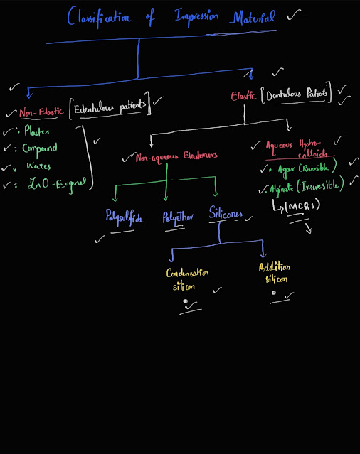
=> Impression material are classified according to their composition, mechanism of setting, mechanical properties and applications.

Classification According to Setting Mechanism

1. Irreversible (Chemical reaction)

Examples includes : Alginate, Plaster of Paris, Silicones ( addition and condensation), Polysulfide, and Polyether.

2. Reversible (Thermally induced physical reaction).

Examples includes : Impression compound and Agar.

Classification according to Mechanical Properties

1. Rigid (inelastic) impression material.

Examples includes: Plaster of Paris, Zinc oxide-eugenol and Impression compound.

2. Elastic impression material.

Examples includes: Alginate, Silicones ( addition and condensation), Polysulfide, and Polyether.
For more click here

Thursday, 2 June 2022

Histology of dentin / Peritubular, Sclerotic and intertubular dentin

Peritubular dentin:

Highly mineralized collar of dentin matrix surrounding the dentinal tubules are called as peritubular dentin.

It is hypermineralized and contain little collagen with large amount of noncollagenous matrix proteins like DSP and DMP1.

Black respresents dentinal tubules and it is surrounded by Peritubular dentin

Sclerotic dentin:

Dentinal tubules which are occluded by calcified matrix are known as sclerotic dentin. 

It is a physiologic process which increase with age by inceased deposition of peritubular dentin. 

It prolongs the vitality of the pulp by preventing the carious lesions reaching to pulp.

Whistish area represents sclerotic dentin

Intertubular dentin:

Dentin which is present between the dentinal tubules are known as intertubular dentin. 

It has high amount of collagen type I having diameter around 50 to 200nm. 

iD= intertubular dentin

For more click here


Dental caries and its classification slideshare ppt

Dental caries It is a multifaceted disease involving teeth host factors of saliva and microflora and the external factors of diet. In it, un...